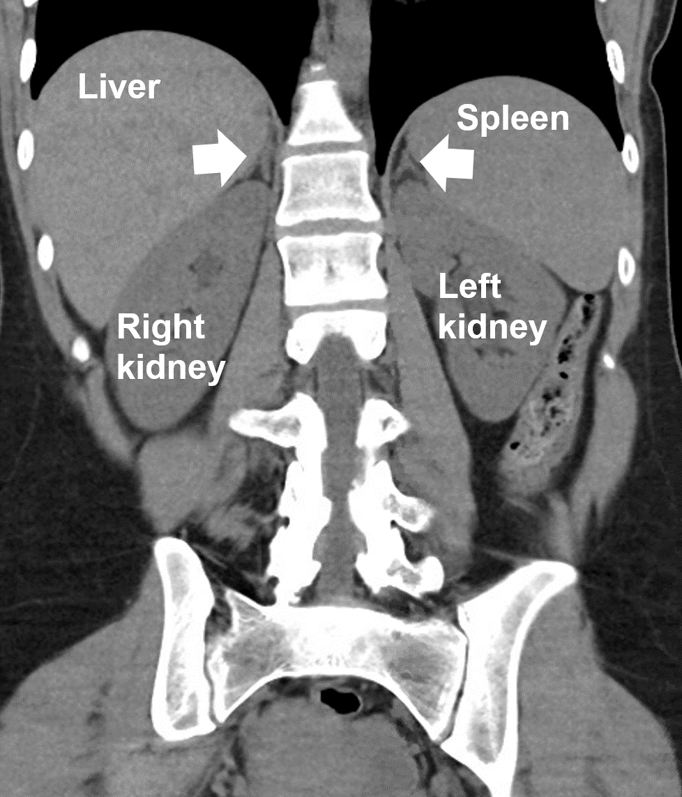
 Adrenal glands are triangle shaped and located on the top of each kidney. CT scan demonstrating the right and left normal adrenal glands (arrows).
Where are Adrenal Glands Located?
Adrenal glands are orange-colored glands located on the top of both kidneys. Adrenal glands are about the size of a fortune cookie and are triangular shaped, sitting on top of the kidneys like hats. Everyone has two adrenal glands that make several important hormones required for healthy life. Although some adrenal conditions are treated with hormones, most adrenal diseases are treated with surgery to remove the adrenal gland or the adrenal tumor that is causing a very specific hormone problem. Adrenal.com has hundreds of pages of information about the adrenal gland organized in easy-to-navigate groups.The Right Adrenal Gland: The adrenal gland on the right side is located on top of the right kidney and is very close to the inferior vena cava (IVC). The IVC is the biggest vein that brings blood back to the heart from the entire body. The right adrenal gland is also very close to the liver. The kidney, IVC and liver are incredibly important structures and it is absolutely crucial that the adrenal surgeon is experienced in dealing with them. If the surgeon is off by just millimeters, the operation can very rapidly end with a tremendously bad outcome.
The Left Adrenal Gland: The left adrenal gland is located on top of the left kidney. It is very close to the splenic artery, which is the major artery that goes to the spleen, and the tail of the pancreas. The pancreas is a very sensitive organ that, when irritated or inflamed, can cause significant sickness and even death. Thus, once again it is exceedingly important that surgery in this area is performed very gently by incredibly experienced surgeons. If the surgeon is off by mere millimeters the operation may end poorly.
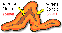 The outer adrenal cortex produces different hormones than the inner adrenal medulla.
FIGURE: A normal adrenal gland demonstrating the adrenal medulla (inner layer), and the adrenal cortex (outer layer). The adrenal glands are shaped like a triangular fortune cookie. And they really are orange in color!
The outer adrenal cortex produces different hormones than the inner adrenal medulla.
FIGURE: A normal adrenal gland demonstrating the adrenal medulla (inner layer), and the adrenal cortex (outer layer). The adrenal glands are shaped like a triangular fortune cookie. And they really are orange in color!
Differences Between the Adrenal Medulla and Cortex.
The adrenal medulla and cortex have completely different functions and produce very different hormones. In fact, when fetuses are only a few weeks old, the adrenal glands form from two different cell types and two different embryonic areas. Inside the Adrenal Gland: The Medulla and Cortex. The adrenal glands are paired (you have two of them) flat glands with a triangular shape, each weighing about 5 grams (about half of a lime). When we cut the adrenal gland in half, we can see with our eyes two distinct regions that are different shades of orange. These two major sections of the adrenal are called the adrenal medulla (inner layer), and the adrenal cortex (outer layer).The Adrenal Medulla: The adrenal medulla comes from the neural crest (i.e. embryological basis similar to your central and peripheral nervous systems). It contains homogenous sheets of cells organized into nests. Cells have large varied nuclei and abundant cytoplasm packed with numerous secretory granules containing catecholamines (fight-or-flight hormones) and other substances specific to chromaffin cells.
The Adrenal Cortex: The adrenal cortex is of mesodermal origin and is derived from the adrenogenital ridge (sorry folks for the technical stuff). The adrenal cortex is organized into three layers, each with a different function:
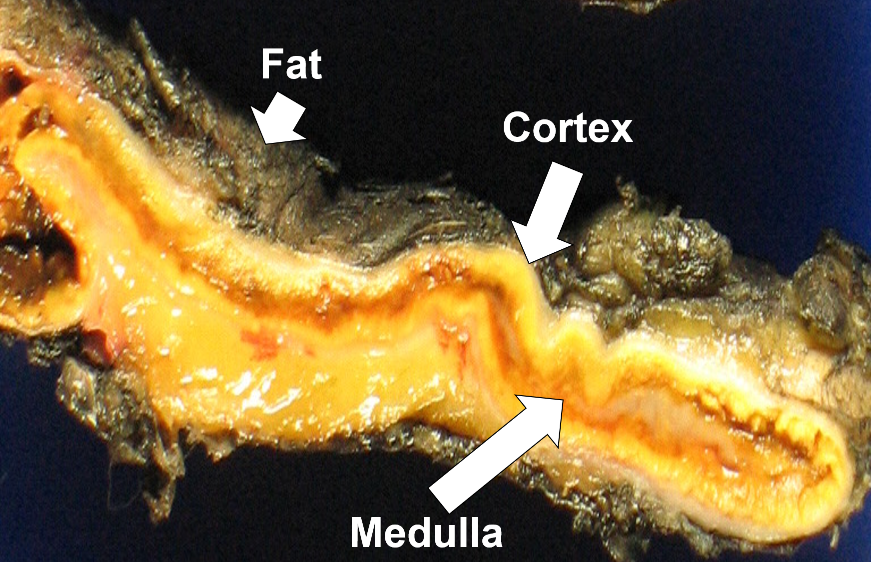 Transection of a portion of normal adrenal tissue displaying the adrenal cortex (yellow), medulla (brownish), and surrounding fat.
- The most superficial layer is the zona glomerulosa, responsible for aldosterone production.
- The middle zone is the zona fasciculata, containing radial columns of lipid-laden (fat-filled) cells that primarily produce cortisol.
- The inner layer, zona reticularis, stores cholesterol for steroidogenesis (the making of steroids) and the secretion of small amounts of sex hormones such as estrogen and testosterone.
Blood Supply to the Adrenal Glands.
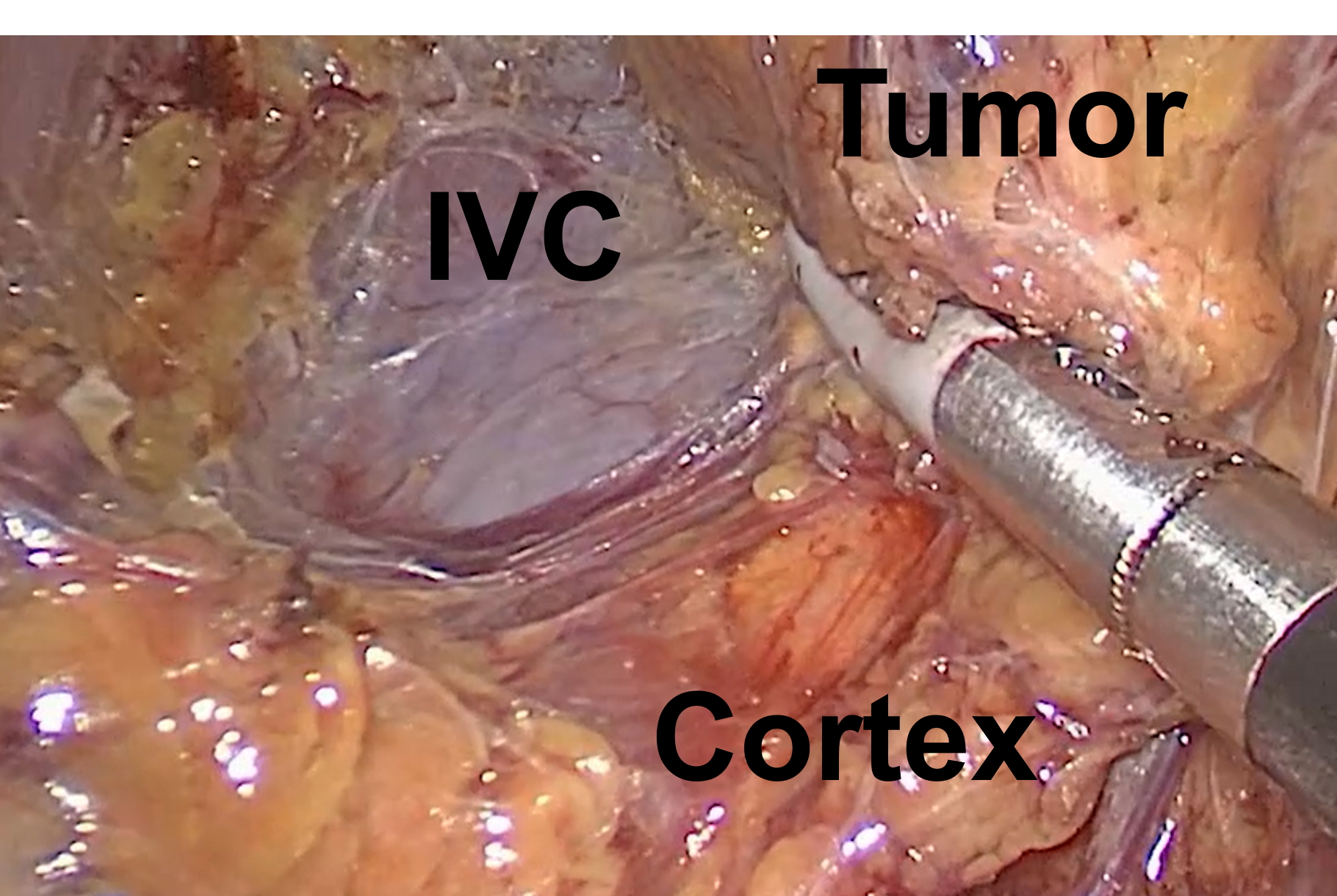 Dissection of the inferior vena cava (IVC), normal adrenal cortex, and the adrenal tumor during a right Mini Back Scope Adrenalectomy (MBSA).
The blood supply to the adrenal is threefold: via the superior adrenal arteries from the inferior phrenic arteries, the middle adrenal from the aorta, and the inferior adrenal artery from the renal arteries. Blood passes from the outer cortex to the inner medulla, and the gland is drained in most cases by a single central vein which empties its blood into the vena cava on the right and the renal vein on the left.
Dissection of the inferior vena cava (IVC), normal adrenal cortex, and the adrenal tumor during a right Mini Back Scope Adrenalectomy (MBSA).
The blood supply to the adrenal is threefold: via the superior adrenal arteries from the inferior phrenic arteries, the middle adrenal from the aorta, and the inferior adrenal artery from the renal arteries. Blood passes from the outer cortex to the inner medulla, and the gland is drained in most cases by a single central vein which empties its blood into the vena cava on the right and the renal vein on the left.