What is the Risk of Cancer in an Adrenal Gland Tumor or Adrenal Mass?
"Is it cancer?" is a very common question we hear at the Carling Adrenal Center. It is important to know that adrenal cancer is quite rare. About 4–7% of adults have an adrenal tumor, more with age, and the large majority are benign. Thus, the risk that you have adrenal cancer is very low based on simple statistics. Adrenocortical cancer and metastatic pheochromocytomas are very uncommon. On the other hand, benign adrenal tumors (which may or may not overproduce hormones) are quite common. The picture is different in people who already have a cancer somewhere else, in whom an adrenal mass may be a metastasis to the adrenal gland.The size of the adrenal mass matters, but size alone does not decide. Very small tumors measuring 1 to 2 cm (less than an inch) are almost never primary adrenal cancers, and the larger the tumor is, the more likely it is to be an adrenal cancer. However, the appearance on the scan, hormone function and growth matter more than size by itself. Small does not always mean safe: a small tumor that looks atypical on CT can still be a concern.
What Size Adrenal Tumor Should be Removed?
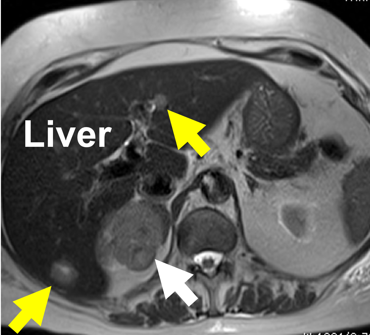 A larger tumor is more likely to be a cancer. This MRI displays an 8 cm right adrenal cancer (white arrow). This patient already had spread (metastasis) to the liver (yellow arrows) at the time of diagnosis.
Tumors over 4 cm (about 1.5 inches) deserve particular attention, and in younger patients we become concerned at a smaller size. If a tumor of this size cannot be shown to be clearly benign on CT, surgery is very likely your best option. A tumor that is uniform and clearly fatty on a non-contrast CT (10 Hounsfield units or less, explained below) is a benign adenoma on imaging and does not need to be removed because of its size alone, although it still needs hormone testing. This is a general guide and there are many exceptions. This is why you need to see a doctor, especially a surgeon, who has seen thousands of these cases and is an expert.
A larger tumor is more likely to be a cancer. This MRI displays an 8 cm right adrenal cancer (white arrow). This patient already had spread (metastasis) to the liver (yellow arrows) at the time of diagnosis.
Tumors over 4 cm (about 1.5 inches) deserve particular attention, and in younger patients we become concerned at a smaller size. If a tumor of this size cannot be shown to be clearly benign on CT, surgery is very likely your best option. A tumor that is uniform and clearly fatty on a non-contrast CT (10 Hounsfield units or less, explained below) is a benign adenoma on imaging and does not need to be removed because of its size alone, although it still needs hormone testing. This is a general guide and there are many exceptions. This is why you need to see a doctor, especially a surgeon, who has seen thousands of these cases and is an expert.
Can a CT Scan Tell if an Adrenal Tumor is Cancer?
Size is not the only criterion in determining whether the tumor is potentially benign or malignant. When we look at the imaging of the adrenal gland, whether it is a CT, MRI, PET scan or other imaging study, we need to have a whole-person approach and evaluate all aspects of the tumor and the patient. We carefully analyze the "imaging phenotype" of the tumor. The imaging phenotype describes all aspects of what the tumor looks like on your scan and helps us determine the likelihood of cancer. It should be noted, this is far from a perfect science and is very dependent upon the experience of your surgeon. In many cases we can never know for sure whether the tumor is cancerous unless we remove it via adrenalectomy.What we look for on scans is a combination of more than a dozen criteria giving us a total impression, called the "imaging phenotype". For instance, we carefully examine the size and the shape of the tumor. That means we will look at whether the borders are smooth or irregular, and whether the margins are clear or unclear. On a CT scan without contrast, it is important to look at the level of attenuation. Essentially, this is a measure of how dense (or fat-containing) the tumor is, measured in Hounsfield units (HU). The lower the Hounsfield units (lipid-rich, i.e. fatty), the more likely it is that the tumor is not a cancer, but rather the more common adrenocortical adenoma. A uniform tumor measuring 10 HU or less is a benign, lipid-rich adenoma. A tumor above 10 HU is "atypical" or indeterminate: many are still benign, but it needs further evaluation, and the higher the density, the more irregular the appearance and the larger the tumor, the greater the concern.
A high-quality non-contrast CT scan is the first and most important scan. When the tumor is not clearly benign on the non-contrast images, an "adrenal protocol" CT with contrast can show how quickly the contrast washes out of the tumor, which can help, but cannot be relied on alone to tell an adenoma from a pheochromocytoma or a malignant tumor. Read more about CT, MRI and PET scans for adrenal tumors.
MRI is rarely the best first test for an adult adrenal tumor. MRI is preferred in pregnancy and in children, where radiation should be avoided, and it is a good alternative when CT contrast cannot be given. If you have only had an MRI, ask whether a non-contrast CT would settle the question.
Other things we look at on the CT scan. We also look to see if there are any areas of the tumor that are dead or necrotic. We look for previous or present bleeding (hemorrhage) into the tumor mass, and we look for calcifications (calcium deposits). The growth rate of the tumor is also very important, so we want to know if the patient has had previous imaging scans of the adrenal glands. A tumor that grows quickly must be assumed to be malignant until proven otherwise. The hormone pattern is another clue: a tumor that makes both cortisol and sex hormones, or symptoms that come on rapidly, raise the suspicion of cancer. Remember that we want to review all your images. Yes, even if you had a scan for unrelated reasons 10 years ago, we want to review it to see what your adrenal glands looked like then.
Should the Adrenal Tumor or Adrenal Mass be Biopsied?
Almost never. A biopsy of an adrenal tumor is rarely needed and can cause significant problems. It can be very dangerous if the patient has a pheochromocytoma, because the biopsy may produce an adrenergic crisis (read about adrenergic crisis in the pheochromocytoma section of this site). For that reason an adrenal tumor should never be biopsied before a pheochromocytoma has been excluded with a blood or urine metanephrine test. If the patient has an adrenocortical carcinoma (adrenal cancer), the biopsy risks spreading tumor cells into the surrounding tissues, which can decrease the chance of curing the patient. Also, a fine needle biopsy cannot reliably distinguish between a benign and a malignant adrenocortical tumor, so it adds risk without answering the question. The main exception is a patient who has another cancer, typically of the lung, kidney, breast or colon, or melanoma, in whom an adrenal metastasis is suspected and the result would change the treatment.We still see adrenal tumors biopsied unnecessarily on a regular basis. If your doctor wants to biopsy your adrenal tumor, ask why, and consider a second opinion from an adrenal specialist.
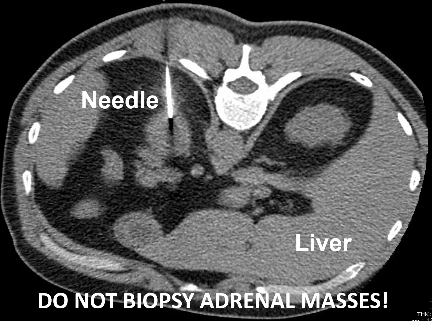 A left adrenocortical cancer undergoing a CT-guided biopsy. This should not be performed when adrenal cancer is suspected.
Now you understand why patients with adrenal tumors should be seen by expert surgeons who have extensive experience with adrenal tumors. It can be somewhat tricky to know in advance of surgery whether the adrenal mass is cancerous or not. Fortunately, the vast majority of adrenal tumors turn out to be benign.
A left adrenocortical cancer undergoing a CT-guided biopsy. This should not be performed when adrenal cancer is suspected.
Now you understand why patients with adrenal tumors should be seen by expert surgeons who have extensive experience with adrenal tumors. It can be somewhat tricky to know in advance of surgery whether the adrenal mass is cancerous or not. Fortunately, the vast majority of adrenal tumors turn out to be benign.
Read about the Mini Back Scope Adrenalectomy (MBSA), which we use for more than 95% of our operations. MBSA suits most tumors; a very large tumor, or one known or strongly suspected to be an adrenal cancer, may need a different approach, and open surgery by an experienced team is the standard for known or suspected adrenocortical carcinoma.
It is important to understand that sometimes surgery to remove an adrenal mass (adrenalectomy) is indicated just to prove that the adrenal tumor is not cancerous. Said differently, sometimes we just don't know if an adrenal mass is a cancer until we get it out. When a tumor cannot be shown to be benign, removing it is, in our experience, usually safer than watching it. Dr. Carling is one of the world's most experienced adrenal surgeons, and he is happy to chat with you and your doctor about your adrenal mass.
Last updated October 2026.