What is the Best Scan for an Adrenal Tumor and Primary Aldosteronism?
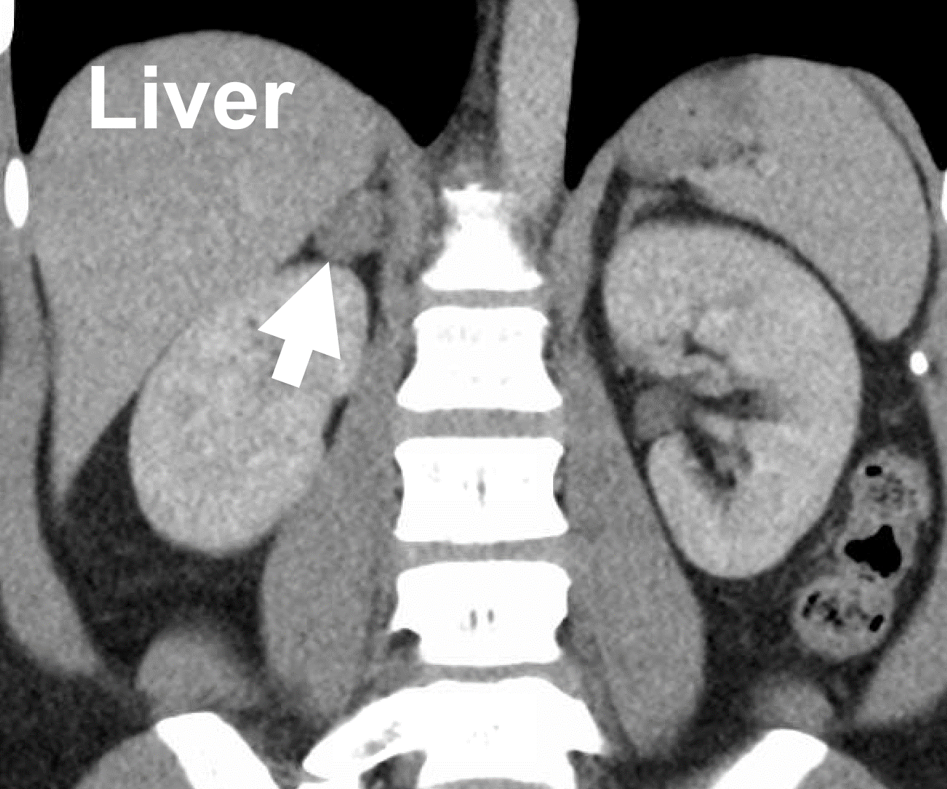 A typical CT scan demonstrating a right aldosterone-producing adrenal adenoma (arrow) causing primary aldosteronism (Conn’s syndrome).
The best first scan for patients with primary aldosteronism is a contrast-enhanced CT scan. On an unenhanced (no contrast) CT, adenomas are usually well-demarcated round or oval lesions, with homogeneous and relatively low density. The addition of contrast helps show the blood vessels better, and thus a CT scan without and with contrast is best. If you are allergic to i.v. contrast, you can have a non-contrast CT scan or an MRI, which is not as good, but better than no scan. In most cases, this scan is the only scan you need. Only occasionally is an MRI helpful in these patients. Dr. Carling has published research on the imaging of aldosterone-producing adenomas (APA). We refer to this type of thin-slice CT scan of the adrenal glands as the adrenal-protocol CT scan.
A typical CT scan demonstrating a right aldosterone-producing adrenal adenoma (arrow) causing primary aldosteronism (Conn’s syndrome).
The best first scan for patients with primary aldosteronism is a contrast-enhanced CT scan. On an unenhanced (no contrast) CT, adenomas are usually well-demarcated round or oval lesions, with homogeneous and relatively low density. The addition of contrast helps show the blood vessels better, and thus a CT scan without and with contrast is best. If you are allergic to i.v. contrast, you can have a non-contrast CT scan or an MRI, which is not as good, but better than no scan. In most cases, this scan is the only scan you need. Only occasionally is an MRI helpful in these patients. Dr. Carling has published research on the imaging of aldosterone-producing adenomas (APA). We refer to this type of thin-slice CT scan of the adrenal glands as the adrenal-protocol CT scan.
A CT scan has limits. It shows the shape of the adrenal glands, not which cells are making aldosterone. Very small aldosterone-producing nodules can be invisible, and a harmless, non-functioning nodule (which becomes more common with age) can point to the wrong side. That is why the scan should be of high quality and read by the surgeon and radiologist together, and why the other adrenal gland must look completely normal before adrenal vein sampling (AVS) is skipped. Newer PET scans that show hormone activity are promising but not yet widely available.
In aldosterone-producing adenomas (APA), there is a correlation between what the tumor looks like on a CT scan, what it looks like under the microscope and which gene is mutated in the tumor (the gene is learned only after surgery). Together with the severity of the disease, this helps us advise who can go straight to surgery and who may need adrenal vein sampling.
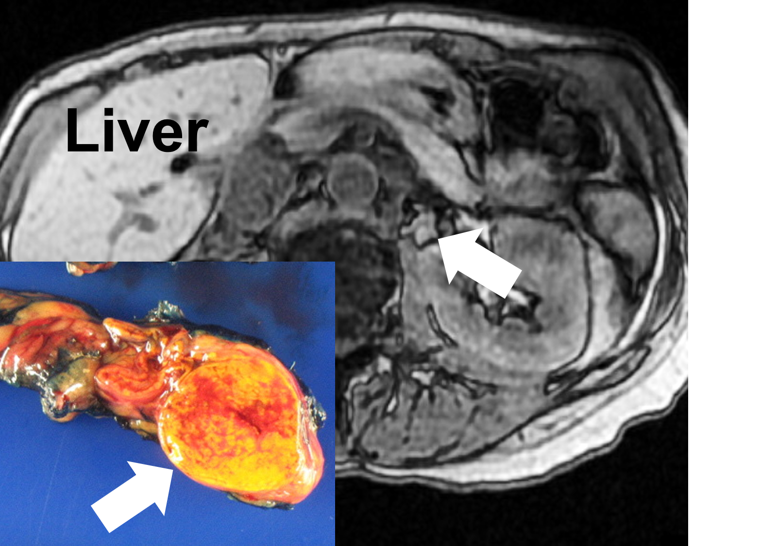 An MRI demonstrating a left aldosterone-producing adenoma (APA; arrow) causing primary aldosteronism (Conn’s syndrome), and after removal via Mini Back Scope Adrenalectomy (MBSA).
Note from the Carling Adrenal Center: If you have primary aldosteronism, we are happy to review your scan alongside your own doctors; we review adrenal scans every day. Do not waste time obtaining unnecessary, expensive scans (for instance, an MRI) or an invasive adrenal vein sampling procedure before your results have been reviewed. Sometimes you need them, but often you do not, and extra scans are often ordered that do not change the plan. In our experience, many patients arrive with scans they did not need; these can confuse the picture and cause patients and doctors unnecessary worry.
An MRI demonstrating a left aldosterone-producing adenoma (APA; arrow) causing primary aldosteronism (Conn’s syndrome), and after removal via Mini Back Scope Adrenalectomy (MBSA).
Note from the Carling Adrenal Center: If you have primary aldosteronism, we are happy to review your scan alongside your own doctors; we review adrenal scans every day. Do not waste time obtaining unnecessary, expensive scans (for instance, an MRI) or an invasive adrenal vein sampling procedure before your results have been reviewed. Sometimes you need them, but often you do not, and extra scans are often ordered that do not change the plan. In our experience, many patients arrive with scans they did not need; these can confuse the picture and cause patients and doctors unnecessary worry.
Adrenal Vein Sampling (AVS) for primary aldosteronism. Adrenal vein sampling is the standard test to distinguish one-sided (unilateral) from two-sided (bilateral) sources of aldosterone. It is an interventional radiology study, and it is not needed for every patient. Patients with severe PA, an unequivocal tumor in one gland on a high-quality CT and a completely normal opposite gland can often go straight to surgery. AVS is most valuable in moderate or severe PA when the CT scan is negative or ambiguous, and it can help when there are tumors in both glands and only a one-sided total adrenalectomy is planned. Through small catheters, blood samples are obtained from each adrenal vein and from the inferior vena cava, sequentially or simultaneously. In experienced centers, both adrenal veins are catheterized and sampled with a technical success rate exceeding 90%. Technical success is directly associated with operator experience, leading to the recommendation that the procedure be performed by one or two operators at an institution to maximize expertise. AVS is invasive and carries a 0.2-2.5% risk of a major complication. Note from the Carling Adrenal Center: Unless your endocrinologist has extensive experience in identifying an expert interventional radiologist to perform adrenal vein sampling (if you need it), call us. We can help you find a team that does this procedure safely and with a high success rate. But remember, many patients with primary aldosteronism do not need adrenal vein sampling.
Warning: Technical Information about Adrenal Vein Sampling: Because the normal adrenal gland produces cortisol in response to ACTH, venous cortisol levels are used as a positive control to determine that each adrenal vein (AV) is adequately sampled. The cortisol level in each adrenal vein is compared to peripheral samples taken from the inferior vena cava. Once it has been shown that both the left and right adrenal veins have been adequately sampled, the next step is to determine whether the results lateralize to one gland (whether the hormone is higher on one side than the other). The aldosterone level (A) in each sample is normalized to the cortisol level. Then, one side is divided by the other to determine the lateralization index (LI). When AVS is done with ACTH (cosyntropin) stimulation, a lateralization index greater than 4 is compatible with a unilateral source of aldosterone; lower cut-offs are used without stimulation. Most centers use the LI to diagnose unilateral PA; suppression of the opposite gland can also support the presence of an aldosterone-producing adenoma (APA). Because 13-27% of patients with PA also make excess cortisol, which can distort these cortisol-corrected ratios, every patient should have a 1-mg dexamethasone suppression test before AVS.
What are the top 10 things to know about adrenal vein sampling for primary aldosteronism (Conn's syndrome)? Read more on our adrenal vein sampling page and on our adrenal surgery blog
If you have an adrenal gland tumor, do not fret. This is all we do, and we can make this easy for you. Send us your laboratory results and your CT scan. Dr. Carling has reviewed the CT scans of thousands of patients with adrenal tumors, and we can usually tell quickly what is going on and what the next step should be. See also our general page on scans for adrenal tumors.
Last updated October 2026.
